


Understanding magnetic tomography: a patient's guide

Magnetic Tomography, often referred to as Magnetic Resonance Imaging (MRI), is a revolutionary diagnostic tool that has transformed the field of medical imaging. It provides detailed images of the organs and tissues within the body, making it an invaluable resource for diagnosing a wide range of conditions, particularly in orthopedic and trauma care.
How Magnetic Tomography Works
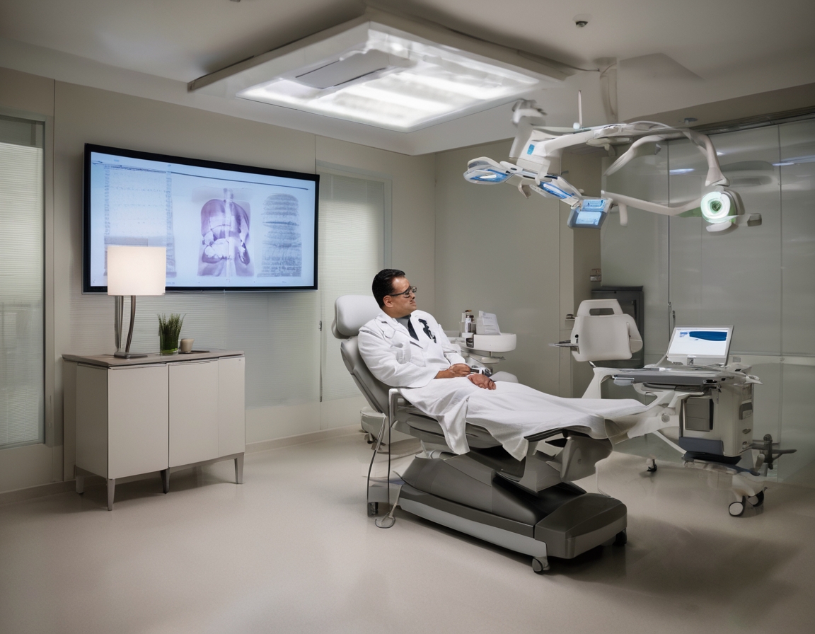
Magnetic Tomography utilizes powerful magnets and radio waves to generate images of the body's internal structures. Unlike X-rays or CT scans, which use ionizing radiation, MRI relies on magnetic fields and radiofrequency pulses to produce detailed images. This makes it a safer option for patients, as it does not expose them to radiation.
The MRI machine is a large, tube-shaped magnet. Patients lie inside the machine during the scan. The magnetic field temporarily realigns hydrogen atoms in the body. Radio waves cause these aligned atoms to produce faint signals, which are used to create cross-sectional MRI images — like slices in a loaf of bread. These images can then be examined from different angles by the radiologist.
Benefits of Magnetic Tomography in Orthopedic and Trauma Care
One of the primary benefits of Magnetic Tomography is its ability to provide highly detailed images of soft tissues, bones, and joints. This precision allows for accurate diagnosis of conditions such as torn ligaments, herniated discs, and other musculoskeletal disorders.
Magnetic Tomography is a non-invasive procedure, meaning it does not require any incisions or injections. This makes it a preferred choice for patients who are apprehensive about invasive diagnostic methods.
Early detection is crucial in managing and treating orthopedic and trauma-related conditions. Magnetic Tomography can identify abnormalities at an early stage, allowing for timely intervention and better treatment outcomes.
Preparing for a Magnetic Tomography Scan
Before undergoing a Magnetic Tomography scan, patients are usually advised to remove any metal objects, such as jewelry or watches, as these can interfere with the magnetic field. Patients should also inform their healthcare provider if they have any metal implants, pacemakers, or other electronic devices.
During the scan, patients will lie on a movable table that slides into the MRI machine. It is important to remain still during the procedure to ensure clear images. The scan is painless, but some patients may feel a bit claustrophobic inside the machine. In such cases, open MRI machines or sedation options may be available.
After the scan, patients can usually resume their normal activities immediately. The images obtained will be analyzed by a radiologist, and the results will be discussed with the patient by their healthcare provider.
Common Conditions Diagnosed with Magnetic Tomography
Magnetic Tomography is particularly useful in diagnosing sports-related injuries, such as ligament tears, muscle strains, and joint abnormalities. It provides detailed images that help in planning effective treatment strategies.
For elderly individuals, Magnetic Tomography can detect degenerative conditions like osteoarthritis and spinal stenosis. Early diagnosis can lead to better management and improved quality of life.
In cases of acute injuries, such as fractures or dislocations, Magnetic Tomography can provide comprehensive images that aid in assessing the extent of the injury and planning appropriate interventions.

Comments (0)